PVCs

Premature Ventricular Contractions

Description

Premature ventricular contractions (PVCs) are beats which are initiated in the ventricles or lower chambers of the heart, prematurely. As opposed to PACs, when the SA node (the natural pacemaker of the heart) gets interrupted, PVCs do not interrupt the SA node. However, with a PVC the ventricles contract, which normally causes the impulse from the atria to be blocked from reaching the ventricles.

PVCs are one of the two most common heart rhythm abnormalities, the other being PACs (premature atrial contractions). They are frequently benign and require no treatment. However, in some cases they may be so frequent (over 15-20/minute) that they may cause the heart to beat inefficiently enough to cause symptoms which may need to be addressed.

PVCs may occur singly or in pairs (generally referred to as couplets), every other beat (bigeminy) or interpolated and may also be described as multiform. Examples of all of these are shown below. (Three or more consecutive PVCs is technically referred to as ventricular tachycardia.) Patients who have these types of rhythm abnormalities may often refer to them as palpitations, skipped beats, hard beats, irregular beats, missing beats or extra beats. They may also complain of feeling dizzy or lightheaded or experience chest pain. Some patients may have no symptoms at all.

Just to reiterate, PVCs are premature beats or beats occurring earlier than they should. Many patients describe them as skipped beats, because when they check their pulse, they don’t feel anything for a moment. However, your heart is not actually skipping or missing a beat. What is happening is that when a beat occurs prematurely, the normal volume of blood has not yet returned from your body from the previous beat. So, even though your heart contracts, not enough blood has returned from the previous beat for it to pump the normal amount of blood. Because of reduced blood being pumped, it may feel like you have skipped a beat, but you have not, although the beat was certainly not as effective as a normal beat.

Patients frequently experience more of these palpitations at night or when they are relaxing. This is because when the natural pacemaker of the heart (the SA node) slows down as it frequently will when you are relaxed, these ectopic (out of the wrong place) foci (point of origins) do not get reset soon enough to stop them.

Examples

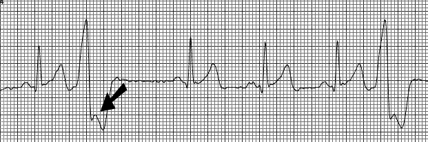
Single PVCs

The above is an example of two isolated PVCs. The heart rate in this example is 70 beats per minute. The first beat, third, fourth and fifth beats are normal, the second and sixth beats are PVCs. There are a number of identifying characteristics which allow us to identify them as PVCs. First, the QRS complex is wide, causing the beat to look strange. (These beats are sometimes referred to euphemistically as FLBs or funny looking beats.) Second, there is a delay after the PVC called a compensatory pause, while the ventricles are waiting for an impulse to reach them from the atria. Third, we can actually see that the atria have contracted and that the SA node has not been reset. If you look carefully in the portion of the QRS complex closest to the bottom of the strip, you will see the waveform rise briefly (arrow), then go down again before rising again. This is actually the P wave, which are the atria contracting. However, this impulse is blocked because the ventricles have just contracted. The next P wave is then conducted normally to the ventricles.

Bigeminal PVCs

The above is an example of three bigeminal PVCs. The heart rate in this example is 83 beats per minute. The first, second, fourth and sixth beats are normal and the third, fifth and seventh are PVCs. PVCs which occur every other beat are called bigeminal PVCs. There may be just a few of these or they may continue for some time.

Multiform PVCs

The above is an example of multiform PVCs (sometimes incorrectly referred to as multifocal PVCs). The heart rate in this example is 63 beats pre minute. The first, third and fifth beats are normal beats (although the observant reader will note that this patient has a bundle branch block) and the second and fourth beats are PVCs, although they are different shapes, thus the term multiform. Generally, they are a different shape because they are originating in two different areas of the ventricles.

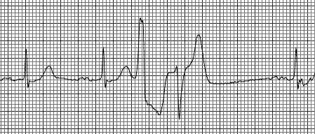
Ventricular Couplets

The above is an example of a ventricular couplet or two consecutive PVCs. The heart rate in this example is 68 beats per minute. In this case they are also multiform PVCs. The first, second and fifth beats are normal and the third and fourth beats are PVCs.

Interpolated PVCs

The above is an example of an interpolated PVC. The heart rate in this example is 68 beats per minute. The first, second, fourth, fifth and sixth beats are normal and the third beat is a PVC. An interpolated PVC is one which is “sandwiched” between two normal beats and therefore does not have a compensatory pause after the PVC. This is because the P wave (which is hidden in the QRS complex) is still conducted to the ventricles and causes them to contract, although there may be a slight delay.