In high degree AV block, two out of every three or more impulses from the atria are blocked by the AV node and fail to reach the ventricles. In third (complete) AV block, all the impulses from the atria are blocked by the AV node. This results in the heart rate being much slower than normal. In certain cases if the subsidiary (backup) pacemakers in the ventricles are absent, the result can be that the ventricles may fail to contract and the heart may stop. These abnormalities may be caused by drug toxicity in which case, modifying the patient’s medications may correct the problem. In other cases, a permanent pacemaker may be required.
Overview
Normal Rhythms
Atrial Arrhythmias
- Atrial Ectopic Rhythm
- Atrial Fibrillation / Flutter
- Junctional Rhythm
- PACs
- PSVT / PAT / SVT
- Sinus Bradycardia
- Sinus Pauses / Asystole
- Sinus Tachycardia
Conduction Disturbances
Ventricular Arrhymias
Miscellaneous Rhythms
Third, High or Complete AV Block
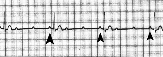
The above EKG shows an example of High Degree AV block. The distinguishing characteristic is that two out of every three P waves are blocked from reaching the ventricles, resulting in an effective heart rate of 34 beats per minute. The arrows point to the P waves that are conducted to the ventricles. The other P waves are blocked by the AV node.
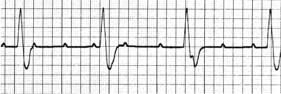
The above EKG shows an example of complete heart block. This rhythm is an idioventricular rhythm. The distinguishing characteristic is that no P waves from the atria are conducted to the ventricles. A careful examination will show that the PR interval is random as a result of there being no relationship between the P wave and the QRS complex. This is also referred to as AV dissociation. The QRS complex in this rhythm actually originates in the ventricles (we know this because there is no relationship between the P wave and the QRS and because the QRS complex is wide, nearly .36 seconds) and the effective heart rate is 33 beats per minute.