Overview
Normal Rhythms
Atrial Arrhythmias
- Atrial Ectopic Rhythm
- Atrial Fibrillation / Flutter
- Junctional Rhythm
- PACs
- PSVT / PAT / SVT
- Sinus Bradycardia
- Sinus Pauses / Asystole
- Sinus Tachycardia
Conduction Disturbances
Ventricular Arrhymias
Miscellaneous Rhythms
Heart Education
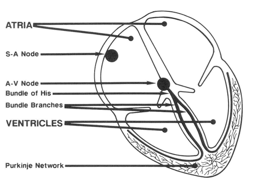
The heart is divided into two primary sections, the atria and the ventricles. Each of these areas is divided into a left and right chamber. The atria are the upper two chambers and the ventricles are the lower two chambers. The wall dividing the left and right sides is the septum. The valves between the atria and ventricles ensure that blood flow is in one direction.
Cardiac contraction is the result of an electrical stimulation of the heart muscle. The normal electrical stimulation starts in special tissue called the SA (sino atrial) node, which is in the right atrium. This is also referred to as the natural pacemaker of the heart. This stimulation or impulse spreads through the atria in a wave like motion. When the atrial contraction reaches the bottom of the right atrium, it stimulates another area of special tissue called the AV (atrial ventricular) node. This takes the impulse from the atria and passes it on to the ventricles by travel through the Bundle of His, Bundle Branches and Purkinje fibers lying in the muscle of the ventricles. When the stimulation reaches the ventricles, a ventricular contraction occurs.
The atria contract just before the ventricles and make sure the ventricles are properly filled before they contract. The atria in effect, give the ventricles a little boost.
The normal heart rate at rest is 60-100 (although we use 50-100 because of how prevalent these slower rates are in the general population) beats per minute and as stated above, the stimulation normally comes from the SA node. However, all cells in the heart can act as pacemakers if the SA node fails. Normally, cells above the ventricles (supraventricular rhythms) will start at 40-60 beats per minute and those in the ventricles at 30-40 beats per minute. Occasionally, an irritable focus will develop which will cause premature beats. If these beats originate in the atria they are called PACs, if originating in the ventricles, they are called PVCs.