You are currently viewing Electrocardiogram (EKG or ECG)

Electrocardiogram (EKG or ECG)

ECG or EKG stands for electrocardiogram and refers to an examination method in which the electrical activity of the heart is measured. The action of the heart is measured with electrodes and recorded in the form of curves. Based on these curves, the doctor can assess whether the heart is functioning properly.

Read here everything about the EKG, how the examination works, and for which patients it is used.

Article overview

 What is an EKG?

An EKG (electrocardiogram or electrocardiography) is an examination method in which the action of the heart, the working cycle of the heart, is measured. The heartbeat is triggered by an electrical signal, which is clocked by the sinus node and propagates through the heart muscle cells. This weak electrical current is measured during the EKG through the electrodes on the extremities or chest wall. Depending on how the EKG electrodes are polarized, a distinction is made between bipolar and unipolar conduction: unipolar conduction refers to a positive electrode with a neutral reference point. In contrast, electrodes with opposite polarity represent the bipolar lead.
The classic EKG is performed with the patient lying down and relaxed and is therefore referred to as the resting EKG. This contrasts with the stress test EKG: Here, the EKG is recorded from the patient during physical exertion – on a treadmill or a bicycle.

Further information: Exercise EKG 

You can read about how an exercise EKG is performed in the article Exercise EKG.
Another special form is the long-term EKG (LZ-EKG), which records the electrical heart activity over 24 hours or even longer.

Further information: Long-term EKG

You can find out about the advantages of the long-term EKG and how the measurement works in the article Long-term EKG.

Excitation generation and conduction in the heart

The heartbeat is generated by a special excitation generation and conduction system: It starts with an electrical impulse in the sinus node, an area in the right atrium of the heart that sets the pace, so to speak. Therefore the sinus node is also called the pacemaker of the heart. The impulse from the sinus node is transmitted to all the muscles in the two atria, which contract and force the blood into the heart chambers.
Then the electrical impulse reaches the AV node, which transfers the electrical stimulus from the atria to the ventricles. The ventricles then contract and transport the blood into the large vessels of the body. While the stimulus is spreading through the ventricles, the excitation in the atria is already receding, the muscles are slackening, and the atria are filling with blood again. After the ventricles have been fully excited, the stimulus here also recedes completely, and the cardiac action starts again from the beginning.

EKG leads

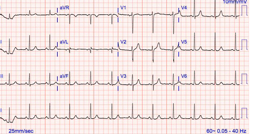
In an extremity EKG, a technician attaches three electrodes to the patient’s body, which is why it is referred to as a 3-lead EKG. Extremity leads include Einthoven bipolar leads (I, II, and III) and Goldberger unipolar leads (aVR, aVL, and aVF). This contrasts with chest wall leads, in which the technician uses six different electrodes and places them on the chest wall (V1-6).
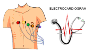
In the classic EKG examination, the chest wall EKG lead and both extremity leads are combined so that a total of twelve electrodes record the electrical stimulus. Therefore, the standard EKG is called a 12-lead EKG.

When do you perform an EKG?

The electrocardiogram provides the physician with information about the rhythm, frequency, excitation, propagation, and regression of the heart. These are frequently altered in the following diseases, among others:
⦁ Heart attack
⦁ Diseases of the coronary vessels (coronary arteries)
⦁ cardiac arrhythmias (atrial fibrillation, ventricular fibrillation, ventricular flutter)
⦁ inflammation of the heart muscle (myocarditis) or pericardium (pericarditis)
⦁ Overdose and poisoning with certain medications (for example, neuroleptics)
⦁ Deficiency or excess of certain minerals (for example, potassium)
⦁ Thickening of the heart wall (right ventricular hypertrophy or left ventricular hypertrophy).
Since some of these conditions are common emergencies, a mobile EKG is available in every ambulance.
Without a prescription, you can buy a Heart Squad Mobile EKG to help prevent problems or monitor your own heart activity.

What do you do during an EKG?

An EKG can be performed both in a doctor’s office and in a hospital or your home. For a resting EKG, the patient lies relaxed on a couch with his or her upper body undressed. Then the doctor or trained technician applies an electrically conductive gel to the EKG electrodes and sticks them to the patient’s skin, depending on the type of lead. Cables connect the electrodes to the EKG machine, which now records the heart’s activity. The examination takes about two minutes. The individual phases of the heart’s action are mapped in characteristic, jagged curves on a paper strip against a time axis. Each deflection represents a specific phase of the heartbeat.

Further information: EKG: Evaluation

What types of spikes and waves there are, how they should look like and what they mean, you can read in the article EKG: Evaluation.

EKG: Extremity leads

In the Einthoven lead, the physician sticks one electrode on each of the patient’s wrists and a reference electrode above the ankle of the left leg. The electrodes are connected bipolar. The following leads are collected:
⦁ Lead I: between the right and left arm; electrical excitation is from right to left.
⦁ Lead II: from the right arm to the left leg
⦁ Lead III: from the left arm to the left leg
In Goldberger conduction, the technician also sticks the electrodes to the wrists and ankles of the left leg, but unlike Einthoven conduction, he connects them unipolar. This results in:
⦁ aVR: right arm
⦁ aVL: left arm
⦁ aVF: left foot

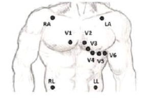
EKG: Chest wall lead according to Wilson.

The technician sticks six electrodes on the patient’s chest, starting just to the right of the sternum and extending to the left lateral chest wall below the armpit. In this way, he obtains leads V1 to V6, each of which is responsible for a specific area of the heart muscle:
⦁ V1 and V2: anterior wall of the cardiac chambers
⦁ V3 and V4: anterior wall of the left ventricle
⦁ V5 and V6: (deep) lateral wall of the left ventricle
If doctors suspect damage to the posterior wall, they stick the electrodes up to the left dorsal area. This results in the additional leads V7, V8 and V9. They represent the electrical activity on the posterior wall of the left ventricle. To better image the right heart leads V3-V6 can also be stuck to the right side of the chest wall in mirror image (V3r-V6r).

What are the risks of an EKG?

The resting and long-term EKG are non-invasive and painless examination methods that are not dangerous for the patient. With the stress EKG, on the other hand, the following problems can occur due to physical exertion, especially in patients with heart conditions:
⦁ Shortness of breath
⦁ Paleness
⦁ Dizziness
⦁ Blood pressure drop or rise
⦁ New onset of arrhythmia (for example, ventricular fibrillation)
⦁ Chest pain or a feeling of tightness or anxiety there (angina pectoris)
⦁ Pulmonary edema (accumulation of fluid in the lungs)
Since the patient is supervised by medical professionals throughout the stress EKG, these problems can be detected in time and the examination can be stopped immediately.

What to expect after an EKG?

After the EKG, the technician removes the electrodes. The contact gel can be easily removed with a paper tissue without leaving any residue. Basically, there are no precautions to be observed. The doctor will explain your findings to you based on the leads and, if necessary, discuss therapy options with you.

Without a prescription, you can buy a Heart Squad Mobile EKG to help prevent problems or monitor your own heart activity.

Leave a Reply